Product Description
In 2020, the Indian orthobiologics market size was valued at $76.2 million, with over 520,000 bone grafting procedures performed every year. The market size is expected to increase at a compound annual growth rate (CAGR) of 12.4% to reach $172.3 million in 2026.
Throughout this medical market research, we analyzed 23 orthobiologics companies across India and used our comprehensive methodology to understand the market sizes, unit sales, company market shares, and to create accurate forecasts.
While this MedSuite report contains all of the Indian Orthopedic Biomaterials market data and analysis, each of the market segments is also available as stand-alone MedCore reports. This allows you to get access to only the market research that you need.
DATA TYPES INCLUDED
- Unit Sales, Average Selling Prices, Market Value & Growth Trends
- Orthobiologics Procedure Volumes
- Market Forecasts Until 2026, and Historical Data to 2016
- Competitive Analysis with Market Shares for Each Segment
- Market Drivers & Limiters for Each Orthopedic Biomaterials Device market
- Recent Mergers & Acquisitions
- Disease Overviews and Demographic Information
- Company Profiles, Product Portfolios and SWOT for Top Competitors
Market Value and Industry Trends
The first bone bank opened in India only in 1997, as compared to 1949 in China. In 2019, the majority of the allograft and DBM allograft supply in India was controlled by two local bone banks, TMH Tissue Bank and Ramaiah Tissue Bank. Other institutions have attempted to open bone banks in the past but have largely been unsuccessful. While the overall trend is positive, the adoption rate is still low, thus limiting the supply side of the market.

Surgical procedures that require the use of orthopedic biomaterials are generally associated with diseases and indications that become more prevalent in the population with an increase in age, such as osteoarthritis of the spine and knee. In 2019, over 6% of India were aged 65 and over; this proportion is projected to grow steadily over the forecast period, as further cohorts of baby boomers turn 65.
Although three-injection products were available in India before single-injection products, the benefits of the latter are typically perceived to outweigh the favorable cost of the former. As a result, single-injection products are expected to continue to cannibalize three-injection products over the forecast period. Due to the premium price of single-injection of products, this will stimulate the total HA viscosupplementation market.
Competitive Analysis

In 2019, the leading competitor in the Indian orthopedic biomaterials market was Sanofi, which was solely attributed to its leading position in the HA viscosupplementation market. The company held the leading position in the single-injection market and the third-leading position in the three-injection market. Sanofi’s portfolio includes the single-injection Synvisc-One® and the three-injection Synvisc® product lines.
Fidia Farmaceutici was the second-leading competitor, which was also attributed to its position in the HA viscosupplementation market. The company held the second-leading position in the single-injection and three-injection markets and the fourth-leading position in the five-injection market. Fidia Farmaceutici’s portfolio includes the single-injection HYALONE® and the three-injection and five-injection HYALGAN® product line.
Segments Covered
Click on each title to view more detailed market segmentation.
- Procedure Volumes for Orthopedic Biomaterials Devices – MedPro – The complete procedural analysis for each segment of the Indian Orthopedic Biomaterials market.
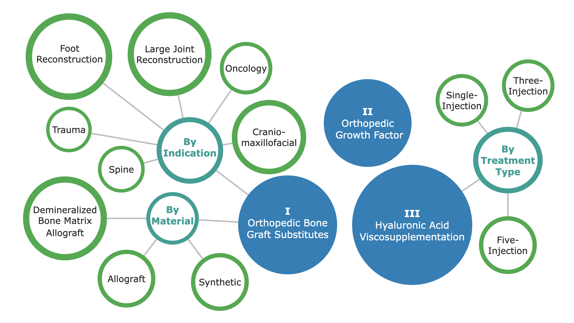
- Orthopedic Bone Graft Substitute Market – MedCore – This market is further segmented by Material Type, including Allograft, Demineralized Bone Matrix Allograft, and Synthetic. Each of these Material Types is segmented by Spine, Trauma, Large Joint Reconstruction, Craniomaxillofacial, and Oncology.
- Orthopedic Growth Factor Market – MedCore – In-depth analysis of the Orthopedic Growth Factor market.
- Hyaluronic Acid Viscosupplementation Market – MedCore – This market is categorized by Treatment Type, including Single-Injection, Three-Injection and Five-Injection.
Detailed Market Segmentation

DON’T SEE THE SEGMENT OR DATA YOU NEED?
Feel free to contact us or send a request by pressing one of the buttons below.
FREE Sample Report
Table of Contents
TABLE OF CONTENTS
TABLE OF CONTENTS I
LIST OF FIGURES VIII
LIST OF CHARTS XV
EXECUTIVE SUMMARY 1
INDIAN ORTHOPEDIC BIOMATERIALS MARKET OVERVIEW 1
COMPETITIVE ANALYSIS 3
MARKET TRENDS 5
MARKET DEVELOPMENTS 6
PROCEDURE NUMBERS 7
MARKETS INCLUDED 8
KEY REPORT UPDATES 10
VERSION HISTORY 10
RESEARCH METHODOLOGY 11
Step 1: Project Initiation & Team Selection 11
Step 2: Prepare Data Systems and Perform Secondary Research 14
Step 3: Preparation for Interviews & Questionnaire Design 16
Step 4: Performing Primary Research 17
Step 5: Research Analysis: Establishing Baseline Estimates 19
Step 6: Market Forecast and Analysis 20
Step 7: Identify Strategic Opportunities 22
Step 8: Final Review and Market Release 23
Step 9: Customer Feedback and Market Monitoring 24
PRODUCT ASSESSMENT 25
2.1 INTRODUCTION 25
2.2 PRODUCT PORTFOLIOS 25
2.2.1 Bone Graft Substitutes 26
2.2.2 Growth Factors 32
2.2.2.1 Other Products 35
2.2.3 Hyaluronic Acid Viscosupplementation (HAV) 37
2.3 REGULATORY ISSUES AND RECALLS 40
2.3.1 Bone Graft Substitutes 40
2.3.1.1 Allografts 40
2.3.1.1.1 MTF 40
2.3.1.2 DBM 40
2.3.1.2.1 AlloSource 40
2.3.1.2.2 SeaSpine 41
2.3.1.2.3 RTI Surgical 41
2.3.1.3 Synthetics 41
2.3.1.3.1 Abyrx 41
2.3.1.3.2 Zimmer Biomet 42
2.3.2 Growth Factors 42
2.3.2.1.1 Medtronic 42
2.3.3 Hyaluronic Acid Viscosupplementation 43
2.3.3.1 Three-injection 43
2.3.3.1.1 Ferring Pharmaceuticals 43
2.4 CLINICAL TRIALS 44
2.4.1 Bone Graft Substitutes 44
2.4.1.1 Allografts 44
2.4.1.1.1 Providence Medical Technology 44
2.4.1.1.2 The University of Texas Health Science 44
2.4.1.1.3 University of Winchester 45
2.4.1.2 BDM 45
2.4.1.2.1 K2M 45
2.4.1.2.2 Zimmer Biomet 46
2.4.1.3 Synthetics 46
2.4.1.3.1 Baxter 46
2.4.1.3.2 Bonesupport 47
2.4.1.3.3 DePuy Synthes 47
2.4.1.3.4 NuVasive 49
2.4.1.3.5 RTI surgical 49
2.4.1.3.6 Sunstar GUIDOR 50
2.4.1.4 Other materials and comparison 51
2.4.1.4.1 Seoul National University Hospital. 51
2.4.1.4.2 Sewon Cellontech 51
2.4.1.4.3 SurgaColl Technologies Limited 52
2.4.1.4.4 University of Colorado 52
2.4.1.4.5 University of Padova 53
2.4.2 Growth Factors 53
2.4.2.1.1 Bioventus 53
2.4.2.1.2 Cerapedics 54
2.4.2.1.3 CGBio 54
2.4.2.1.4 Isto 55
2.4.2.1.5 Medtronic 55
2.4.2.1.6 NuVasive 57
2.4.2.1.7 Wright 57
2.4.2.1.8 Others 58
2.4.3 Hyaluronic Acid Viscosupplementation 59
2.4.3.1.1 Anika Therapeutics 59
2.4.3.1.2 Bioventus 60
2.4.3.1.3 Cairo University 61
2.4.3.1.4 Ferring Pharmaceuticals 61
2.4.3.1.5 Federal University of Minas Gerais 62
2.4.3.1.6 Fidia Pharma USA Inc. 62
2.4.3.1.7 Istituto Ortopedico Rizzoli 63
2.4.3.1.8 University Hospital 63
2.4.3.1.9 Universidade Nova de Lisboa 64
INDIAN ORTHOPEDIC BIOMATERIALS MARKET OVERVIEW 65
3.1 INTRODUCTION 65
3.1.1 Bone Graft Substitutes 65
3.1.2 Growth Factors 65
3.1.3 Hyaluronic Acid (HA) Viscosupplementation 66
3.2 CURRENCY EXCHANGE RATE 67
3.3 MARKET OVERVIEW AND TREND ANALYSIS 68
3.4 DRIVERS AND LIMITERS 76
3.4.1 Market Drivers 76
3.4.2 Market Limiters 76
3.5 COMPETITIVE MARKET SHARE ANALYSIS 78
3.6 MERGERS AND ACQUISITIONS 83
3.7 COMPANY PROFILES 85
3.7.1 Anika Therapeutics 85
3.7.2 Bioventus 86
3.7.3 DePuy Synthes 87
3.7.4 Ferring Pharmaceuticals 88
3.7.5 Fidia Pharmaceuticals 89
3.7.6 Genzyme (Sanofi Group) 90
3.7.7 Harvest Technologies (Terumo BCT) 91
3.7.8 Integra LifeSciences 92
3.7.9 Medtronic 93
3.7.10 Musculoskeletal Transplant Foundation (MTF) 95
3.7.11 NuVasive 96
3.7.12 Orthofix 97
3.7.13 RTI Surgical 98
3.7.14 Stryker 99
3.7.15 Vericel Corporation (formerly Aastrom Bioscience) 100
3.7.16 Zimmer Biomet 101
3.8 SWOT ANALYSIS 103
3.8.1 Anika Therapeutics 103
3.8.2 Bioventus 105
3.8.3 DePuy Synthes 107
3.8.4 Ferring Pharmaceuticals 108
3.8.5 Fidia Pharmaceuticals 109
3.8.7 Harvest Technologies (Terumo BCT) 111
3.8.8 Integra LifeSciences 112
3.8.9 Medtronic 113
3.8.10 Musculoskeletal Transplant Foundation (MTF) 114
3.8.11 NuVasive 115
3.8.12 Orthofix 116
3.8.14 Stryker 118
3.8.15 Vericel Corporation (formerly Aastrom Bioscience) 119
3.8.16 Zimmer Biomet 120
PROCEDURE NUMBERS 122
4.1 INTRODUCTION 122
4.1.1 Units per Procedure by Indication 123
4.2 PROCEDURES 124
4.2.1 Orthopedic Biomaterial Procedures by Segment 124
4.2.2 Orthopedic Bone Grafting Procedures 126
4.2.2.1 Orthopedic Bone Grafting Procedures by Material 126
4.2.2.1.1 Autograft Procedures by Indication 128
4.2.2.1.2 Allograft Procedures by Indication 131
4.2.2.1.3 DBM Allograft Procedures by Indication 134
4.2.2.1.4 Synthetic Procedures by Indication 137
4.2.2.2 Orthopedic Bone Grafting Procedures by Indication 140
4.2.3 Orthopedic Growth Factor Procedures 143
4.2.4 Hyaluronic Acid Supplementation Procedures 145
4.2.4.1 Hyaluronic Acid Supplementation Procedures by Injection Cycle 145
ORTHOPEDIC BONE GRAFT SUBSTITUTE MARKET 147
5.1 INTRODUCTION 147
5.2 MARKET OVERVIEW 148
5.2.1 Orthopedic Bone Graft Substitute Market by Material 148
5.2.2 Orthopedic Bone Graft Substitute Market by Indication 153
5.3 MARKET ANALYSIS AND FORECAST 159
5.3.1 Total Orthopedic Bone Graft Substitute Market 159
5.3.2 Orthopedic Bone Graft Substitute Market by Material 161
5.3.2.1 Allograft Bone Graft Substitute Market 161
5.3.2.1.1 Allograft Bone Graft Substitute Market by Indication 163
5.3.2.2 Demineralized Bone Matrix Allograft Bone Graft Substitute Market 177
5.3.2.2.1 Demineralized Bone Matrix Allograft Bone Graft Substitute Market by Indication 179
5.3.2.3 Synthetic Bone Graft Substitute Market 193
5.3.2.3.1 Synthetic Bone Graft Substitute Market by Indication 195
5.3.3 Orthopedic Bone Graft Substitute Market by Indication 209
5.3.3.1 Spine Bone Graft Substitute Market 209
5.3.3.2 Cervical Spine Bone Graft Substitute Market 211
5.3.3.3 Thoracolumbar Spine Bone Graft Substitute Market 213
5.3.3.4 Trauma Bone Graft Substitute Market 215
5.3.3.5 Non-Union Trauma Bone Graft Substitute Market 217
5.3.3.6 Fresh Fracture Trauma Bone Graft Substitute Market 219
5.3.3.7 Large Joint Reconstruction Bone Graft Substitute Market 221
5.3.3.8 Hip Reconstruction Bone Graft Substitute Market 223
5.3.3.9 Knee Reconstruction Bone Graft Substitute Market 225
5.3.3.10 Foot Reconstruction Bone Graft Substitute Market 227
5.3.3.11 Craniomaxillofacial Bone Graft Substitute Market 229
5.3.3.12 Oncology Bone Graft Substitute Market 231
5.4 DRIVERS AND LIMITERS 233
5.4.1 Market Drivers 233
5.4.2 Market Limiters 233
5.5 COMPETITIVE MARKET SHARE ANALYSIS 235
ORTHOPEDIC GROWTH FACTOR MARKET 238
6.1 INTRODUCTION 238
6.1.1 BMP-2 (Medtronic) 238
6.2 MARKET ANALYSIS AND FORECAST 240
6.3 DRIVERS AND LIMITERS 242
6.3.1 Market Drivers 242
6.3.2 Market Limiters 242
6.4 COMPETITIVE MARKET SHARE ANALYSIS 244
HYALURONIC ACID VISCOSUPPLEMENTATION MARKET 245
7.1 INTRODUCTION 245
7.1.1 Benefits of Viscosupplementation 246
7.1.2 Synovial Fluid 246
7.2 MARKET OVERVIEW 247
7.3 MARKET ANALYSIS AND FORECAST 253
7.3.1 Total Hyaluronic Acid Viscosupplementation Market 253
7.3.2 Single-Injection Hyaluronic Acid Viscosupplementation Market 255
7.3.3 Three-Injection Hyaluronic Acid Viscosupplementation Market 257
7.3.4 Five-Injection Hyaluronic Acid Viscosupplementation Market 259
7.4 DRIVERS AND LIMITERS 261
7.4.1 Market Drivers 261
7.4.2 Market Limiters 261
7.5 COMPETITIVE MARKET SHARE ANALYSIS 263
ABBREVIATIONS 267
Companies Included
The Orthobiologics Market Report Suite | India | 2020 - 2026 | MedSuite includes analysis on the following companies currently active in this market:
• Alkem Laboratories
• LG Chem
• Anika Therapeutics
• Medtronic
• Basic Healthcare
• MEI
• Bharat Biotech
• NovaBone
• Biocomposites
• Sanofi
• Bioventus
• Sun Pharmaceutical
• BONESUPPORT
• Surgiwear
• DePuy Synthes
• Universe Surgical
• Dr. Reddy’s Laboratories
• Virchow Biotech
• Ferring Pharmaceuticals
• Wright Medical
• Fidia Farmaceutici
• Zimmer Biomet
• IFGL Bio Ceramics
Chart List
LIST OF CHARTS
Chart 1 1: Orthopedic Biomaterials Market by Segment, India, 2016 – 2026 2
Chart 1 2: Orthopedic Biomaterials Market Overview, India, 2019 & 2026 2
Chart 3 1: Orthopedic Biomaterials Market by Segment, India, 2016 – 2026 71
Chart 3 2: Orthopedic Biomaterials Market Breakdown, India, 2019 72
Chart 3 3: Orthopedic Biomaterials Market Breakdown, India, 2026 73
Chart 3 4: Growth Rates by Segment, Orthopedic Biomaterials Market, India, 2017 – 2026 75
Chart 3 5: Leading Competitors, Orthopedic Biomaterials Market, India, 2019 82
Chart 4 1: Orthopedic Biomaterials Market by Segment, India, 2016 – 2026 125
Chart 4 2: Orthopedic Bone Grafting Procedures by Material, India, 2016 – 2026 127
Chart 4 3: Autograft Procedures by Indication, India, 2016 – 2026 130
Chart 4 4: Allograft Procedures by Indication, India, 2016 – 2026 133
Chart 4 5: DBM Allograft Procedures by Indication, India, 2016 – 2026 136
Chart 4 6: Synthetic Procedures by Indication, India, 2016 – 2026 139
Chart 4 7: Orthopedic Bone Grafting Procedures by Indication, India, 2016 – 2026 142
Chart 4 8: Orthopedic Growth Factor Procedures, India, 2016 – 2026 144
Chart 4 9: Hyaluronic Acid Viscosupplementation Procedures by Injection Cycle, India, 2016 – 2026 146
Chart 5 1: Orthopedic Bone Graft Substitute Market by Material, India, 2016 – 2026 150
Chart 5 2: Orthopedic Bone Graft Substitute Market Breakdown by Material, India, 2019 151
Chart 5 3: Orthopedic Bone Graft Substitute Market Breakdown by Material, India, 2026 152
Chart 5 4: Orthopedic Bone Graft Substitute Market by Indication, India, 2016 – 2026 156
Chart 5 5: Orthopedic Bone Graft Substitute Market Breakdown by Indication, India, 2019 157
Chart 5 6: Orthopedic Bone Graft Substitute Market Breakdown by Indication, India, 2026 158
Chart 5 7: Total Orthopedic Bone Graft Substitute Market, India, 2016 – 2026 160
Chart 5 8: Allograft Bone Graft Substitute Market, India, 2016 – 2026 162
Chart 5 9: Allograft Bone Graft Substitute Market by Indication, India, 2016 – 2026 164
Chart 5 10: Demineralized Bone Matrix Allograft Bone Graft Substitute Market, India, 2016 – 2026 178
Chart 5 11: Demineralized Bone Matrix Allograft Bone Graft Substitute Market by Indication, India, 2016 – 2026 180
Chart 5 12: Synthetic Bone Graft Substitute Market, India, 2016 – 2026 194
Chart 5 13: Synthetic Bone Graft Substitute Market by Indication, India, 2016 – 2026 196
Chart 5 14: Spine Bone Graft Substitute Market, India, 2016 – 2026 210
Chart 5 15: Cervical Spine Bone Graft Substitute Market, India, 2016 – 2026 212
Chart 5 16: Thoracolumbar Spine Bone Graft Substitute Market, India, 2016 – 2026 214
Chart 5 17: Trauma Bone Graft Substitute Market, India, 2016 – 2026 216
Chart 5 18: Non-Union Trauma Bone Graft Substitute Market, India, 2016 – 2026 218
Chart 5 19: Fresh Fracture Trauma Bone Graft Substitute Market, India, 2016 – 2026 220
Chart 5 20: Large Joint Reconstruction Bone Graft Substitute Market, India, 2016 – 2026 222
Chart 5 21: Hip Reconstruction Bone Graft Substitute Market, India, 2016 – 2026 224
Chart 5 22: Knee Reconstruction Bone Graft Substitute Market, India, 2016 – 2026 226
Chart 5 23: Foot Reconstruction Bone Graft Substitute Market, India, 2016 – 2026 228
Chart 5 24: Craniomaxillofacial Bone Graft Substitute Market, India, 2016 – 2026 230
Chart 5 25: Oncology Bone Graft Substitute Market, India, 2016 – 2026 232
Chart 5 26: Leading Competitors, Orthopedic Bone Graft Substitute Market, India, 2019 237
Chart 6 1: Orthopedic Growth Factor Market, India, 2016 – 2026 241
Chart 7 1: Hyaluronic Acid Viscosupplementation Market by Segment, India, 2016 – 2026 250
Chart 7 2: Hyaluronic Acid Viscosupplementation Market Breakdown, India, 2019 251
Chart 7 3: Hyaluronic Acid Viscosupplementation Market Breakdown, India, 2026 252
Chart 7 4: Total Hyaluronic Acid Viscosupplementation Market, India, 2016 – 2026 254
Chart 7 5: Single-Injection Hyaluronic Acid Viscosupplementation Market, India, 2016 – 2026 256
Chart 7 6: Three-Injection Hyaluronic Acid Viscosupplementation Market, India, 2016 – 2026 258
Chart 7 7: Five-Injection Hyaluronic Acid Viscosupplementation Market, India, 2016 – 2026 260
Chart 7 8: Leading Competitors, Hyaluronic Acid Viscosupplementation Market, India, 2019 266
Figure List
LIST OF FIGURES
Figure 1 1: Orthopedic Biomaterials Market Share Ranking by Segment, India, 2019 3
Figure 1 2: Companies Researched in this Report 4
Figure 1 3: Factors Impacting the Orthopedic Biomaterials Market by Segment, India 5
Figure 1 4: Recent Events in the Orthopedic Biomaterials Market, India, 2017 – 2019 6
Figure 1 5: Orthopedic Biomaterials Procedures Covered, India 7
Figure 1 6: Orthopedic Biomaterials Markets Covered (1 of 2) 8
Figure 1 7: Orthopedic Biomaterials Markets Covered (2 of 2) 9
Figure 1 8: Key Report Updates 10
Figure 1 9: Version History 10
Figure 2 1: Bone Graft Substitutes Products by Company (1 of 4) 28
Figure 2 2: Bone Graft Substitutes Products by Company (2 of 4) 29
Figure 2 3: Bone Graft Substitutes Products by Company (3 of 4) 30
Figure 2 4: Bone Graft Substitutes Products by Company (4 of 4) 31
Figure 2 5: Growth Factor Products by Company 36
Figure 2 6: Hyaluronic Acid Viscosupplementation by Products by Company 39
Figure 2 7: Class 2 Device Recall Musculoskeletal Transplant Foundation Allofix Insertion Kit 40
Figure 2 8: Class 2 Device Recall AlloFuse DBM Putty 5cc 40
Figure 2 9: Class 2 Device Recall Accell Evo3c Demineralized Bone Matrix Putty 41
Figure 2 10: Class 2 Device Recall RTI Biologics BioSet IC RT Paste 2 cc 41
Figure 2 11: Class 2 Device Recall Hemostatic Bone Putty 41
Figure 2 12: Class 2 Device Recall Endobon Xenograft Granules 42
Figure 2 13: Class 2 Device Recall Endobon Xenograft Granules 42
Figure 2 14: Class 2 Device Recall INFUSE Bone Graft X SMALL KIT 42
Figure 2 15: Class 3 Device Recall Euflexxa (1 sodium hyaluronate) 43
Figure 2 16: Evaluation of DTRAX Graft in Patients with Cervical Degenerative Disc Disease 44
Figure 2 17: Ridge Preservation Using FDBA and a Collagen Wound Dressing in Molar Sites. 44
Figure 2 18: Assessing Physical Activity Levels of Patients Following HTO. 45
Figure 2 19: Evaluation of Fusion Rate Using K2M VESUVIUS® Demineralized Fibers with K2M EVEREST® Spinal System 45
Figure 2 20: Evaluation of Zimmer Puros® Allograft vs. Creos™ Allograft for Alveolar Ridge Preservation 46
Figure 2 21: Synthetic Bone Graft Substitute vs. Autologous Spongiosa in Revision Anterior Cruciate Ligament Reconstruction 46
Figure 2 22: Cerament Treatment of Fracture Defects (CERTiFy) 47
Figure 2 23: Comparison of Bioactive Glass and Beta-Tricalcium Phosphate as Bone Graft Substitute (BAGvsTCP) 47
Figure 2 24: Evaluation of Fusion Rate of Anterior Cervical Discectomy and Fusion (ACDF) Using Cervios ChronOs™ and Bonion™ 48
Figure 2 25: AttraX® Putty vs. Autograft in XLIF® 49
Figure 2 26: Comparison of nanOss Bioactive with Autograft and Bone Marrow Aspirate to Autograft in the Posterolateral Spine 49
Figure 2 27: Assessment of nanOss Bioactive 3D in the Posterolateral Spine 50
Figure 2 28: Assessment of Ridge Preservation Using Moldable Beta-tricalcium Phosphate Bone Grafting System 50
Figure 2 29: Outcome Comparison of Allograft and Synthetic Bone Substitute in High Tibial Osteotomy 51
Figure 2 30: Efficacy and Safety of SurgiFill™ on Spinal Fusion 51
Figure 2 31: Assessment of HydroxyColl Bone Graft Substitute in High Tibial Osteotomy Wedge Grafting. (HColl_HTO) 52
Figure 2 32: Outcomes of the Evans Calcaneal Lengthening Based on Bone Grafting Material 52
Figure 2 33: Deproteinized Bovine Bone in Alveolar Bone Critical Size Defect (>2cm) Secondary to Cyst Removal 53
Figure 2 34: A Prospective Study of Instrumented, Posterolateral Lumbar Fusions (PLF) With OsteoAMP® 53
Figure 2 35: The Clinical Effect of i-FACTOR® Versus Allograft in Non-instrumented Posterolateral Spondylodesis Operation 54
Figure 2 36: Clinical Study of Injectable Ceramics Bone Graft Substitute Containing rhBMP-2 54
Figure 2 37: Prospective Study of Safety and Efficacy of InQu® Bone Graft Extender in Lumbar Interbody Fusion Surgery (Intebody) 55
Figure 2 38: A Study of INFUSE Bone Graft (BMP-2) in the Treatment of Tibial Pseudarthrosis in Neurofibromatosis Type 1 55
Figure 2 39: Clinical Study of INFUSE® Bone Graft Compared to Autogenous Bone Graft for Vertical Ridge Augmentation 56
Figure 2 40: Parallel Study Between BMP-2 and Autologous Bone Graft After Ilizarow Treatment 56
Figure 2 41: RCT of AttraX® Putty vs. Autograft in Instrumented Posterolateral Spinal Fusion (AxA) 57
Figure 2 42: Long-term Safety and Effectiveness of AUGMENT® Bone Graft Compared to Autologous Bone Graft 57
Figure 2 43: rhBMP-2 vs Autologous Bone Grafting for the Treatment of Non-union of the Docking Site in Tibial Bone Transport 58
Figure 2 44: Evaluation of Radiculitis Following Use of Bone Morphogenetic Protein-2 for Interbody Arthrodesis in Spinal Surgery 58
Figure 2 45: Study of Cingal™ for the Relief of Knee Osteoarthritis Compared to Triamcinolone Hexacetonide at 39 Weeks Follow-Up (Cingal17-02) 59
Figure 2 46: HyaloFAST Trial for Repair of Articular Cartilage in the Knee (FastTRACK) 59
Figure 2 47: Effectiveness of Two Hyaluronic Acids in Osteoarthritis of the Knee 60
Figure 2 48: The Effect of Topical Application of Hyaluronic Acid on Immediate Dental Implant 61
Figure 2 49: To Look at the Characteristics of Synovial Fluid and Cartilage Matrix in Osteoarthritic Knees After Hyaluronic Acid Injection 61
Figure 2 50: Use of Hyaluronic Acid as a Therapeutic Strategy for Bone Repair in Humans 62
Figure 2 51: Two Weekly Intra-articular Hyaluronan Knee Injections, Given One Week Apart, of HYMOVIS Combined With a Physical Exercise Program (PEP) Compared to PEP Alone, in a Relatively Young, Active Population of Subjects With Patellofemoral Osteoarthritis (PFOA) and/or Tibiofemoral Osteoarthritis (TFOA) 62
Figure 2 52: Comparative Assessment of Viscosupplementation With Polynucleotides and Hyaluronic Acid (PNHA1401) 63
Figure 2 53: Trial Comparing Botulin Toxin Versus Hyaluronic Acid by Intra-articular Injection (GOTOX) 63
Figure 2 54: Trial to Assess the Structural Effect and Long-term Symptomatic Relief of Intra-articular Injections of HA (ViscOA) 64
Figure 3 1: Currency Exchange Rate, 2019 67
Figure 3 2: Orthopedic Biomaterials Market by Segment, India, 2016 – 2026 (US$M) 69
Figure 3 3: Orthopedic Biomaterials Market by Segment, India, 2016 – 2026 (IN₹M) 70
Figure 3 4: Orthopedic Biomaterials Market Growth by Segment, India, 2016 – 2026 74
Figure 3 5: Drivers and Limiters, Orthopedic Biomaterials Market, India, 2019 77
Figure 3 6: Leading Competitors, Orthopedic Biomaterials Market, India, 2019 81
Figure 3 7: SWOT Analysis, Anika Therapeutics (1 of 2) 103
Figure 3 8: SWOT Analysis, Anika Therapeutics (2 of 2) 104
Figure 3 9: SWOT Analysis, Bioventus (1 of 2) 105
Figure 3 10: SWOT Analysis, Bioventus (2 of 2) 106
Figure 3 11: SWOT Analysis, DePuy Synthes 107
Figure 3 12: SWOT Analysis, Ferring Pharmaceuticals 108
Figure 3 13: SWOT Analysis, Fidia Pharmaceuticals 109
Figure 3 14: SWOT Analysis, Genzyme (Sanofi) 110
Figure 3 15: SWOT Analysis, Harvest Technologies 111
Figure 3 16: SWOT Analysis, Integra LifeSciences 112
Figure 3 17: SWOT Analysis, Medtronic 113
Figure 3 18: SWOT Analysis, MTF 114
Figure 3 19: SWOT Analysis, NuVasive 115
Figure 3 20: SWOT Analysis, Orthofix 116
Figure 3 21: SWOT Analysis, RTI Surgical 117
Figure 3 22: SWOT Analysis, Stryker 118
Figure 3 23: SWOT Analysis, Vericel Corporation 119
Figure 3 24: SWOT Analysis, Zimmer Biomet (1 of 2) 120
Figure 3 25: SWOT Analysis, Zimmer Biomet (2 of 2) 121
Figure 4 1: Units per Procedure by Indication, Bone Graft Substitute Market, India, 2016– 2026 123
Figure 4 2: Orthopedic Biomaterials Procedures by Segment, India, 2016 – 2026 124
Figure 4 3: Orthopedic Bone Grafting Procedures by Material, India, 2016 – 2026 126
Figure 4 4: Autograft Procedures by Indication, India, 2016 – 2026 (1 of 2) 128
Figure 4 5: Autograft Procedures by Indication, India, 2016 – 2026 (2 of 2) 129
Figure 4 6: Allograft Procedures by Indication, India, 2016 – 2026 (1 of 2) 131
Figure 4 7: Allograft Procedures by Indication, India, 2016 – 2026 (2 of 2) 132
Figure 4 8: DBM Allograft Procedures by Indication, India, 2016 – 2026 (1 of 2) 134
Figure 4 9: DBM Allograft Procedures by Indication, India, 2016 – 2026 (2 of 2) 135
Figure 4 10: Synthetic Procedures by Indication, India, 2016 – 2026 (1 of 2) 137
Figure 4 11: Synthetic Procedures by Indication, India, 2016 – 2026 (2 of 2) 138
Figure 4 12: Orthopedic Bone Grafting Procedures by Indication, India, 2016 – 2026 (1 of 2) 140
Figure 4 13: Orthopedic Bone Grafting Procedures by Indication, India, 2016 – 2026 (2 of 2) 141
Figure 4 14: Orthopedic Growth Factor Procedures, India, 2016 – 2026 143
Figure 4 15: Hyaluronic Acid Viscosupplementation Procedures by Injection Cycle, India, 2016 – 2026 145
Figure 5 1: Orthopedic Bone Graft Substitute Market by Material, India, 2016 – 2026 (US$M) 148
Figure 5 2: Orthopedic Bone Graft Substitute Market by Material, India, 2016 – 2026 (IN₹M) 149
Figure 5 3: Orthopedic Bone Graft Substitute Market by Indication, India, 2016 – 2026 (US$M) 154
Figure 5 4: Orthopedic Bone Graft Substitute Market by Indication, India, 2016 – 2026 (IN₹M) 155
Figure 5 5: Total Orthopedic Bone Graft Substitute Market, India, 2016 – 2026 159
Figure 5 6: Allograft Bone Graft Substitute Market, India, 2016 – 2026 161
Figure 5 7: Allograft Bone Graft Substitute Market by Indication, India, 2016 – 2026 (US$M) 163
Figure 5 8: Spine Allograft Market, India, 2016 – 2026 165
Figure 5 9: Cervical Spine Allograft Market, India, 2016 – 2026 166
Figure 5 10: Thoracolumbar Spine Allograft Market, India, 2016 – 2026 167
Figure 5 11: Trauma Allograft Market, India, 2016 – 2026 168
Figure 5 12: Non-Union Trauma Allograft Market, India, 2016 – 2026 169
Figure 5 13: Fresh Fracture Trauma Allograft Market, India, 2016 – 2026 170
Figure 5 14: Large Joint Reconstruction Allograft Market, India, 2016 – 2026 171
Figure 5 15: Hip Reconstruction Allograft Market, India, 2016 – 2026 172
Figure 5 16: Knee Reconstruction Allograft Market, India, 2016 – 2026 173
Figure 5 17: Foot Reconstruction Allograft Market, India, 2016 – 2026 174
Figure 5 18: Craniomaxillofacial Allograft Market, India, 2016 – 2026 175
Figure 5 19: Oncology Allograft Market, India, 2016 – 2026 176
Figure 5 20: Demineralized Bone Matrix Allograft Bone Graft Substitute Market, India, 2016 – 2026 177
Figure 5 21: Demineralized Bone Matrix Allograft Bone Graft Substitute Market by Indication, India, 2016 – 2026 (US$M) 179
Figure 5 22: Spine DBM Allograft Market, India, 2016 – 2026 181
Figure 5 23: Cervical Spine DBM Allograft Market, India, 2016 – 2026 182
Figure 5 24: Thoracolumbar Spine DBM Allograft Market, India, 2016 – 2026 183
Figure 5 25: Trauma DBM Allograft Market, India, 2016 – 2026 184
Figure 5 26: Non-Union Trauma DBM Allograft Market, India, 2016 – 2026 185
Figure 5 27: Fresh Fracture Trauma DBM Allograft Market, India, 2016 – 2026 186
Figure 5 28: Large Joint Reconstruction DBM Allograft Market, India, 2016 – 2026 187
Figure 5 29: Hip Reconstruction DBM Allograft Market, India, 2016 – 2026 188
Figure 5 30: Knee Reconstruction DBM Allograft Market, India, 2016 – 2026 189
Figure 5 31: Foot Reconstruction DBM Allograft Market, India, 2016 – 2026 190
Figure 5 32: Craniomaxillofacial DBM Allograft Market, India, 2016 – 2026 191
Figure 5 33: Oncology DBM Allograft Market, India, 2016 – 2026 192
Figure 5 34: Synthetic Bone Graft Substitute Market, India, 2016 – 2026 193
Figure 5 35: Synthetic Bone Graft Substitute Market by Indication, India, 2016 – 2026 (US$M) 195
Figure 5 36: Spine Synthetic Market, India, 2016 – 2026 197
Figure 5 37: Cervical Spine Synthetic Market, India, 2016 – 2026 198
Figure 5 38: Thoracolumbar Spine Synthetic Market, India, 2016 – 2026 199
Figure 5 39: Trauma Synthetic Market, India, 2016 – 2026 200
Figure 5 40: Non-Union Trauma Synthetic Market, India, 2016 – 2026 201
Figure 5 41: Fresh Fracture Trauma Synthetic Market, India, 2016 – 2026 202
Figure 5 42: Large Joint Reconstruction Synthetic Market, India, 2016 – 2026 203
Figure 5 43: Hip Reconstruction Synthetic Market, India, 2016 – 2026 204
Figure 5 44: Knee Reconstruction Synthetic Market, India, 2016 – 2026 205
Figure 5 45: Foot Reconstruction Synthetic Market, India, 2016 – 2026 206
Figure 5 46: Craniomaxillofacial Synthetic Market, India, 2016 – 2026 207
Figure 5 47: Oncology Synthetic Market, India, 2016 – 2026 208
Figure 5 48: Spine Bone Graft Substitute Market, India, 2016 – 2026 209
Figure 5 49: Cervical Spine Bone Graft Substitute Market, India, 2016 – 2026 211
Figure 5 50: Thoracolumbar Spine Bone Graft Substitute Market, India, 2016 – 2026 213
Figure 5 51: Trauma Bone Graft Substitute Market, India, 2016 – 2026 215
Figure 5 52: Non-Union Trauma Bone Graft Substitute Market, India, 2016 – 2026 217
Figure 5 53: Fresh Fracture Trauma Bone Graft Substitute Market, India, 2016 – 2026 219
Figure 5 54: Large Joint Reconstruction Bone Graft Substitute Market, India, 2016 – 2026 221
Figure 5 55: Hip Reconstruction Bone Graft Substitute Market, India, 2016 – 2026 223
Figure 5 56: Knee Reconstruction Bone Graft Substitute Market, India, 2016 – 2026 225
Figure 5 57: Foot Reconstruction Bone Graft Substitute Market, India, 2016 – 2026 227
Figure 5 58: Craniomaxillofacial Bone Graft Substitute Market, India, 2016 – 2026 229
Figure 5 59: Oncology Bone Graft Substitute Market, India, 2016 – 2026 231
Figure 5 60: Drivers and Limiters, Orthopedic Bone Graft Substitute Market, India, 2019 234
Figure 5 61: Leading Competitors, Orthopedic Bone Graft Substitute Market, India, 2019 236
Figure 6 1: Orthopedic Growth Factor Market, India, 2016 – 2026 240
Figure 6 2: Drivers and Limiters, Orthopedic Growth Factor Market, India, 2019 243
Figure 6 3: Leading Competitors, Orthopedic Growth Factor Market, India, 2019 244
Figure 7 1: Hyaluronic Acid Viscosupplementation Market by Segment, India, 2016 – 2026 (US$M) 248
Figure 7 2: Hyaluronic Acid Viscosupplementation Market by Segment, India, 2016 – 2026 (IN₹M) 249
Figure 7 3: Total Hyaluronic Acid Viscosupplementation Market, India, 2016 – 2026 253
Figure 7 4: Single-Injection Hyaluronic Acid Viscosupplementation Market, India, 2016 – 2026 255
Figure 7 5: Three-Injection Hyaluronic Acid Viscosupplementation Market, India, 2016 – 2026 257
Figure 7 6: Five-Injection Hyaluronic Acid Viscosupplementation Market, India, 2016 – 2026 259
Figure 7 7: Drivers and Limiters, Hyaluronic Acid Viscosupplementation Market, India, 2019 262
Figure 7 8: Leading Competitors, Hyaluronic Acid Viscosupplementation Market, India, 2019 265
iData’s 9-Step Research Methodology
Our reports follow an in-depth 9-step methodology which focuses on the following research systems:
- Original primary research that consists of the most up-to-date market data
- Strong foundation of quantitative and qualitative research
- Focused on the needs and strategic challenges of the industry participants
Step 1: Project Initiation & Team Selection During this preliminary investigation, all staff members involved in the industry discusses the topic in detail.
Step 2: Prepare Data Systems and Perform Secondary Research The first task of the research team is to prepare for the data collection process: Filing systems and relational databases are developed as needed.
Step 3: Preparation for Interviews & Questionnaire Design The core of all iData research reports is primary market research. Interviews with industry insiders represent the single most reliable way to obtain accurate, current data about market conditions, trends, threats and opportunities.
Step 4: Performing Primary Research At this stage, interviews are performed using contacts and information acquired in the secondary research phase.
Step 5: Research Analysis: Establishing Baseline Estimates Following the completion of the primary research phase, the collected information must be synthesized into an accurate view of the market status. The most important question is the current state of the market.
Step 6: Market Forecast and Analysis iData Research uses a proprietary method to combine statistical data and opinions of industry experts to forecast future market values.
Step 7: Identify Strategic Opportunities iData analysts identify in broad terms why some companies are gaining or losing share within a given market segment.
Step 8: Final Review and Market Release An integral part of the iData research methodology is a built-in philosophy of quality control and continuing improvement is integral to the iData philosophy.
Step 9: Customer Feedback and Market Monitoring iData philosophy of continuous improvement requires that reports and consulting projects be monitored after release for customer feedback and market accuracy.
Click Here to Read More About Our Methodology